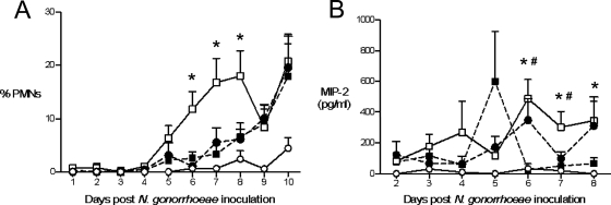
FIG. 4.
A greater PMN influx occurs in coinfected mice. (A) Significantly more PMNs migrate into the lower genital tracts of mice coinfected with N. gonorrhoeae and C. muridarum (white squares, solid line) compared to mice infected with N. gonorrhoeae (black circles, dashed line) or C. muridarum (black squares, dashed line) alone, or uninfected mice (white circles, solid line) on days 6, 7, and 8 after N. gonorrhoeae inoculation (*, P < 0.05). The percent PMNs is defined as the number of PMNs counted/100 vaginal cells observed, including squamous and nucleated epithelial cells and leukocytes. The results shown are from three combined experiments (n = 30 to 32 mice per group). (B) Levels of PMN attracting chemokine MIP-2 are greatest on days 6 to 8 in mice coinfected with N. gonorrhoeae and C. muridarum (white squares, solid line) compared to mice infected with either N. gonorrhoeae (black circles, dashed line) or C. muridarum (black squares, dashed line) alone or uninfected mice (white circles, solid line) on days 6 to 8 (*, P < 0.05, coinfected versus uninfected; #, P < 0.05, coinfected versus C. muridarum alone). The results from a single experiment are shown (n = 4 to 5 mice per group). The high average level of MIP-2 detected on day 5 in C. muridarum-infected mice is due to one mouse in this group having 1,843 pg of MIP-2/ml. MIP-2 concentrations ranged from 60 to 646 pg/ml for the other four mice within this group.

