FIG. 4.
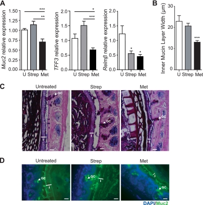
Metronidazole treatment alters goblet cell function and reduces the production of the inner mucus layer. (A) Quantitative RT-PCR results of Muc2, TFF3, and Relmβ expression in the distal colon. Results are averaged from three independent experiments (n = 3 to 6 mice per group). U, untreated mice; for Muc2, ***, P = 0.0008, and **, P = 0.0017; for TFF3, ***, P < 0.0001, and *, P = 0.02; for Relmβ, Strep *, P = 0.03, and Met *, P = 0.01. (B) Quantification of inner mucin layer thickness. Distal colon sections were fixed in methanol-Carnoy's fixative, embedded in paraffin, and stained with AB/PAS to visualize and quantify the inner mucus layer. The inner mucus width was determined by an average of 4 measurements per field with 4 fields counted per tissue section. Results are averaged from two independent experiments (n = 3 mice per group). U, untreated mice; ***, P = 0.0003. (C) AB/PAS-stained methanol-Carnoy's fixative-fixed distal colon sections showing the inner mucin layer (white arrowheads). i, inner mucin layer; GC, goblet cell. Original magnification, ×400. Bars, 20 μm. (D) Representative immunostaining for the inner mucin layer using an antibody that recognizes murine Muc2 (green) with DAPI (blue) as a counterstain. The inner mucin layer is thinner in metronidazole-treated C57BL/6 mice. i, inner mucin layer; GC, goblet cell. Original magnification, ×400. Bars, 20 μm.

