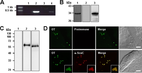
FIG. 2.
Expression of ScaC by O. tsutsugamushi. (A) RT-PCR of scaC mRNA from L929 cells infected with O. tsutsugamushi. scaC DNA was amplified from total RNA (without reverse transcription) isolated from infected cells (lane 1), from genomic DNA (lane 2), from cDNA from uninfected cells (lane 3), and from cDNA from infected cells (lane 4). (B) Specificity of anti-ScaC antiserum. Purified His-tagged ScaC33-232 protein was resolved by SDS-PAGE, stained with Coomassie blue (lane 1), and immunoblotted with rabbit preimmune serum (lane 2) or anti-ScaC serum (lane 3). (C) Immunoblot analysis of total O. tsutsugamushi proteins by using rabbit preimmune serum (lane 1) or anti-ScaC serum (lane 2). Anti-ScaC serum detected a protein with a molecular mass of approximately 60 kDa. Preimmune serum did not react with the bacterial lysate. Immunoblotting using anti-TAS56 was performed as a control (lane 3). (D) Immunofluorescence confocal microscopy using preimmune serum or anti-ScaC serum (α-ScaC) showed ScaC in the O. tsutsugamushi-infected L929 cells. The left-hand panels show bacteria (O. tsutsugamushi [OT]) stained with pooled serum from scrub typhus patients. Magnified images are shown in the lower panels (inset boxes). Scale bars, 5 μm.

