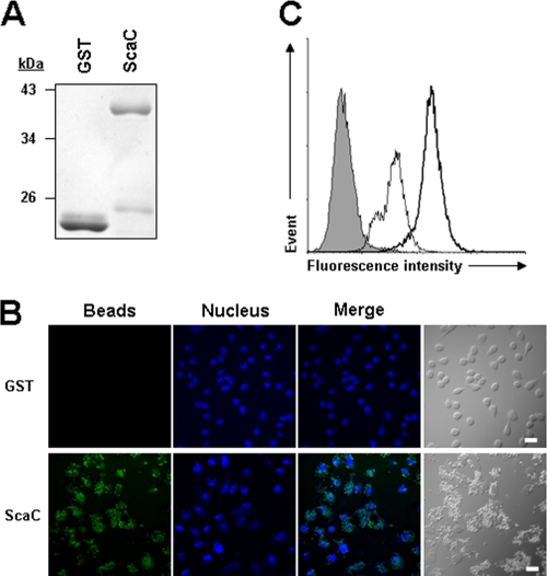
FIG. 4.
Adhesion of ScaC-coated microbeads to HeLa cells. (A) Purity of recombinant GST or GST-ScaC (ScaC) protein for the conjugation with fluorescent microbeads was assessed by SDS-PAGE followed by Coomassie blue staining. (B) Cells were incubated with fluorescent microbeads coated with GST or GST-ScaC (ScaC) for 1 h, washed extensively, and fixed. Cell-bound microbeads (green) were visualized by fluorescence microscopy after staining of cell nuclei (blue). Scale bars, 10 μm. (C) Relative binding of the microbeads coated with GST (thin line) or GST-ScaC (thick line) to HeLa cells was quantified directly using fluorescence-activated cell sorter (FACS) analysis. The gray histogram represents unbound cells (cells not incubated with microbeads).

