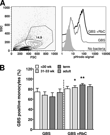
FIG. 4.
Nonopsonic and opsonic phagocytosis of GBS by neonatal and adult monocytes. CBMC or PBMC were incubated for 1 h with pHrodo dye-labeled HKGBS (1 × 10⁸ CFU/ml) with our without RbC. (A) Left panel, representative forward-scatter (FSC) versus side-scatter (SSC) FACS plot showing inclusion gate for monocytes; right panel, representative histograms showing pHrodo-specific fluorescence in monocytes without bacteria or with GBS plus or minus RbC. (B) Percentage of monocytes positive for phagocytosed GBS (means ± SEM). n = 11 for the <30-week group, 15 for the 31- to 33-week and term groups, and 18 for adults. *, P < 0.0151; **, P < 0.0084; Wilcoxon signed rank test comparing phagocytosis of GBS with and without RbC.

