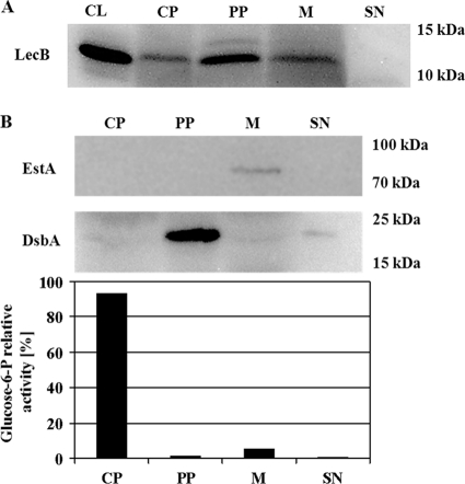
FIG. 1.
Subcellular localization of LecB in P. aeruginosa PAO1 and controls to validate the cellular fractionation procedure. (A) Western blot analysis of cell fractions from P. aeruginosa PAO1. Cells were grown at 37°C on NB agar plates for 48 h, subcellular fractions were prepared as described in the text and separated by SDS-gel electrophoresis, and LecB was detected after transfer onto PVDF membranes using a LecB-specific antiserum. (B) Fractionation controls. The same cell fractions were analyzed using an EstA- and a DsbA-specific antiserum and by the distribution of relative glucose-6-phosphate dehydrogenase activities. The percentages of relative enzyme activities present in cytoplasm, periplasm, membrane, and culture supernatant are given. CL, cell lysate; CP, cytoplasm; PP, periplasm; M, membrane; SN, culture supernatant.

