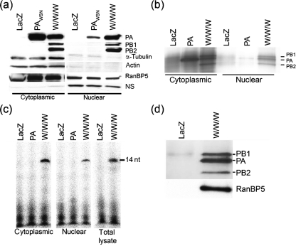
Fig. 1.
Subcellular distribution and activity of purified, adenovirus-expressed WSN polymerase. (a) A549 cells were transduced with rAd5-PAWSN-TAP alone or with a combination of rAd5-PAWSN-TAP, -PB1WSN and -PB2WSN (W/W/W). Nuclear and cytoplasmic extracts were analysed by immunoblotting using PA-, PB1- or PB2-specific antibodies. The effectiveness of the subcellular fractionation method was confirmed by probing against the cytoplasmic proteins, actin and β-tubulin as well as against RanBP5 (which is found both in the cytoplasm and nucleus). ns, non-specific protein band detected when probing against PB1/2. Cells transduced with an adenovirus vector expressing LacZ served as a negative control. Twofold more cell equivalents were loaded for the nuclear extracts than the cytoplasmic extracts. (b) IgG-purified nuclear and cytoplasmic extracts were analysed by silver staining prior to (c) comparison with total cell lysate in an ApG-primed transcription assay (1 h at 30 °C). Extracts from equivalent numbers of transduced A549 cells were used. (d) IgG-purified extracts from total cellular lysates were analysed by silver staining (upper panel) and immunoblotting against RanBP5 (lower panel). The positions of PA, PB1 and PB2 are shown on the right.

