FIG. 3.
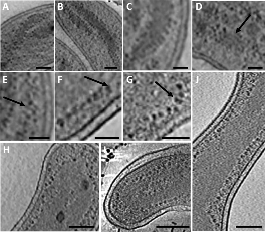
Well-separated nucleoid spirals and ribosome distribution at the edge of the nucleoid in MreB2-mTFP (panels A to G, I, and J) and wild-type (H) cells. (A and B) Ribosomes (dark dots) line the edge of the nucleoid (medium-gray density). (C, D, and E) Nucleoid protrusions, 10 nm wide, or parallel ribbons (arrow) are decorated by ribosomes. (F and G) Pairs or strings of ribosomes (arrow) line the nucleoid edge. Nucleoid-associated ribosomes in a wild-type cell (H) and in two other MreB2-mTFP mutant cells (I and J). Bars, 100 nm (panel A), 75 nm (panels B, D, and E), 50 nm (panels C, F, and G), and 200 nm (panels H to J).

