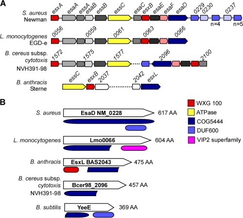
FIG. 1.
Schematic of Ess loci and EsaD-like proteins in Gram-positive bacteria. (A) Comparison of the S. aureus Ess (type VII-like) secretion locus with L. monocytogenes and “Bacillus cytotoxicus” ess loci, as well as the B. anthracis ess gene cluster (16). Genes showing sequence homology are depicted in the same color. (B) Domain organization of S. aureus EsaD and protein homologues showing the conserved COG5444 domain. With the exception of B. subtilis YeeE, all COG5444 proteins lie within an Ess-like locus. The colors of some conserved proteins and domains indicate WXG100 proteins (red), FtsK SpoIIIE-like ATPases (yellow), EsaD and COG5444 (dark blue), proteins with a DUF600 domain (light blue), and VIP2 (pink).

