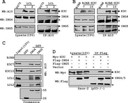
FIG. 1.
EBNA3C forms a p53-independent complex with both ING4 and ING5. BJAB cells and two BJAB cell stable clones expressing EBNA3C (E3C7 and 10) (A) and DG75 cells and two LCL clones (LCL1 and 2) (B) were subjected to IP with EBNA3C specific antibody (A10). Samples were resolved by SDS-PAGE and detected by Western blotting (WB) for the indicated proteins by stripping and reprobing the same membrane. (C) Either purified GST or GST-ING4 or -ING5 beads were incubated with cell lysates, and EBNA3C was detected by WB with A10. Coomassie staining of GST-labeled proteins is shown in the bottom panel. Numbers at the right of the bottom blot are molecular masses (in kilodaltons). (D) Saos-2 (p53−/−) cells were cotransfected with myc-EBNA3C in the presence of either the vector control, flag-tagged ING4, or flag-tagged ING5, as indicated. At 36 h posttransfection, cells were harvested, lysed, and immunoprecipitated with flag antibody. Samples were subjected to WB with the indicated antibodies. The asterisks indicate the immunoglobulin bands.

