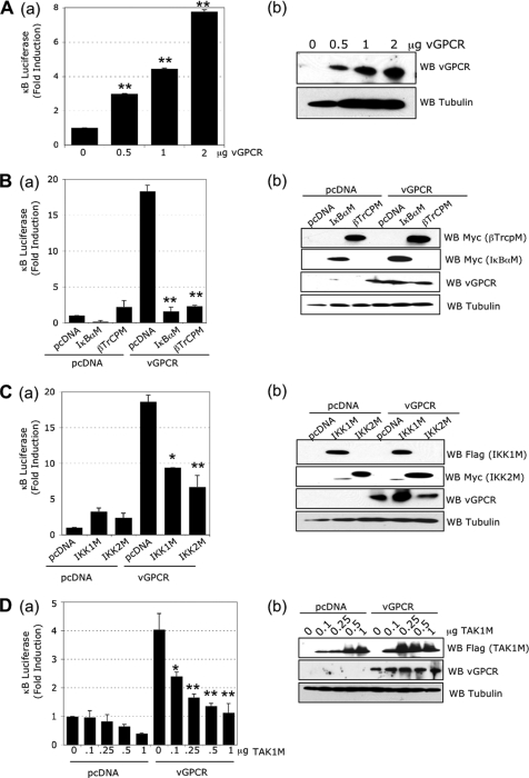
FIG. 1.
The classical NF-κB pathway is induced by vGPCR. (A) (a) vGPCR induces NF-κB activation. HEK293T cells were transfected with pcDNA or increasing quantities of vGPCR expression plasmids, 1 μg of κB-luciferase construct, and 0.5 μg of β-Gal reporter construct. The luciferase and β-galactosidase activities were measured in triplicate using Steady-glo and Beta-glo Promega kits, respectively. (b) Western blots (WB) using lysates from transfected cells with the indicated antibodies. (B) (a) NF-κB activation induced by vGPCR is blocked by IκBα and βTrCP mutants. HEK293T cells were transfected with 1 μg of pcDNA or vGPCR expression plasmids in the presence or absence of 1 μg of IκBα mutant (IκBαM) or βTrCP mutant (βTrCPM) expression plasmid, along with κB-luciferase and β-Gal reporter constructs. (b) Western blots from lysates of transfected cells using the indicated antibodies. (C) (a) NF-κB activation induced by vGPCR is blocked by IKK1 and IKK2 mutants. HEK293T cells were transfected with 1 μg of pcDNA or vGPCR expression plasmids in the presence or absence of 1 μg of IKK1 or IKK2 mutant expression plasmids (IKK1M and IKK2M, respectively), along with κB-luciferase and β-Gal reporter constructs. (b) Western blots from transfected cell lysates using the indicated antibodies. (D) (a) NF-κB activation induced by vGPCR is blocked by a TAK1 mutant. HEK293T cells were transfected with 1 μg of pcDNA or vGPCR expression plasmids, an increasing quantity of TAK1M expression plasmid, along with κB-luciferase and β-Gal reporter constructs. Statistics compare vGPCR conditions with the corresponding pcDNA conditions at the same quantity of TAK1M plasmid. (b) Western blots from transfected cell lysates using the indicated antibodies. The bar graphs represent the mean and standard deviation (SD) of fold induction (n = 3). *, P < 0.05; **, P < 0.01 compared to the pcDNA control within the vGPCR group (unless stated otherwise).

