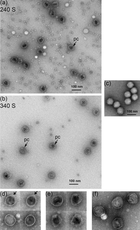
FIG. 6.
EM images of negatively stained Syn5 procapsids. (a) Micrograph of particles in the 240 S sucrose gradient fraction showing procapsid particles (pc, arrow) filled with protein density (most likely the scaffolding protein, making them impermeable to stain) and empty, stain-permeable capsids. (b) Procapsid particles (pc, arrows) in the 340 S fraction were similar to those in the 240 S peak, with diameters of about 50 nm. Empty shells were observed more rarely. (c) Syn5 mature virions for size and shape comparison. (d) Panel of enlarged procapsidlike particles found in both peaks. The arrows indicate areas of higher density at the coat wall. (e) Enlarged empty capsids present in both fractions. (f) Icosahedral capsids found in the 340 S peak. All samples were negatively stained with 1% uranyl acetate and observed at a magnification of ×120,000.

