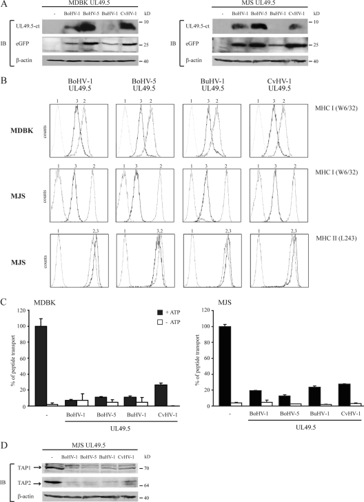
FIG. 2.
The UL49.5 proteins of BoHV-5, BuHV-1, and CvHV-1 inhibit peptide transport and mediate the degradation of TAP1 and TAP2. (A) Lysates derived from control and UL49.5-expressing MDBK and MJS cells were stained for GFP and UL49.5 by SDS-PAGE and immunoblotting (IB) with specific antibodies. The β-actin signal was used as a loading control. (B) Surface expression of MHC-I (MDBK and MJS) and MHC-II (MJS) molecules was assessed by flow cytometry on untransduced cells (graph 2) and on cells expressing the UL49.5 homologs of BoHV-1, BoHV-5, BuHV-1, and CvHV-1 (graph 3) using the indicated antibodies. Graph 1, background staining in the presence of secondary antibody only. (C) Transport activity of TAP was analyzed in MDBK and MJS expressing the UL49.5 homologs. Peptide transport was evaluated in the presence of ATP (▪) or EDTA (□). (D) Steady-state levels of TAP1 and TAP2 in MJS cells were determined using specific antibodies. The β-actin signal was used as a loading control.

