FIG. 4.
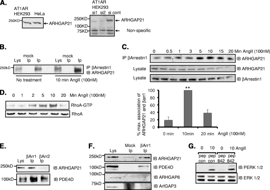
Dynamic interaction between β-arrestin 1 and ARHGAP21 in AT1AR HEK 293 cells. (A) (Left) Cell lysates from AT1AR HEK 293 cells and HeLa cells were blotted for ARHGAP21. (Right) The specificity of the antibody was verified using siRNA silencing of ARHGAP21. cont, control. (B) Immunoprecipitates (IP) of β-arrestin 1 from AT1AR HEK 293 cells were blotted (IB) for ARHGAP21 before or after treatment with angiotensin II (AngII) (100 nM). (C) (Top) Immunoprecipitates of β-arrestin 1 from AT1AR HEK 293 cells were blotted for ARHGAP21 following a time course of treatment with angiotensin II (100 nM). (Center panels) Equal amounts of ARHGAP21 and β-arrestin 1 in inputs. (Bottom) Bar chart showing quantifications of the upper trace. max., maximum; βarr1, β-arrestin 1. (D) RhoA activation in AT1AR HEK 293 cells as measured by a GST-Rhotekin pulldown assay following a time course of angiotensin II treatment. (E) Immunoprecipitates of β-arrestin 1 or β-arrestin 2 from AT1AR HEK 293 cells were blotted for ARHGAP21 and PDE4D. (F) Immunoprecipitates of β-arrestin 1 from AT1AR HEK 293 cells were blotted for ARHGAP21, PDE4D, ARHGAP6, and ArfGAP3. (G) Determination of total and phospho-ERK 1 and 2 in lysates of AT1AR HEK 293 cells following angiotensin II treatment after pretreatment with peptide 842 (pep 842) or a control peptide (pep con). PERK, phospho-ERK.

