
FIG. 3.
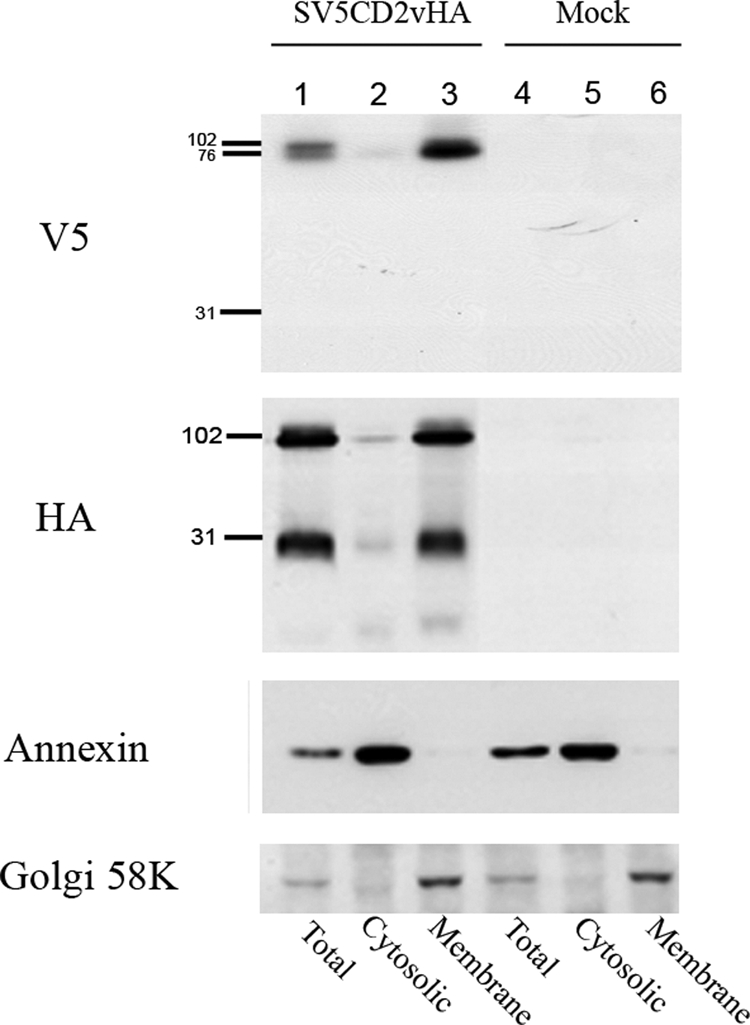
Western blot showing the localization of CD2v protein fragments in membrane or cytosolic cell fractions in ASFV-infected cells. Vero cells were transfected with plasmids expressing SV5CD2vHA and infected with the ASFV BA71V isolate. At 18 h postinfection, the cells were lysed and separated into cytosolic or membrane fractions. Lanes 1, 2, and 3, extracts from cells expressing the SV5CD2vHA protein; lanes 4, 5, and 6, extracts from mock-transfected cells. Lanes 1 and 4 show total cell extracts prepared by lysis in RIPA buffer, and lanes 2 to 6 show extracts fractionated into the cytosolic (lanes 2 and 5) and membrane (lanes 3 and 6) fractions. Extracts were separated by SDS-PAGE and blotted onto membranes. The blots were probed as indicated on the left with anti-V5-HRP, anti-HA-HRP, anti-annexin followed by anti-mouse-HRP, and anti-Golgi 58K protein followed by anti-mouse-HRP. Bound antibodies were detected by ECL.