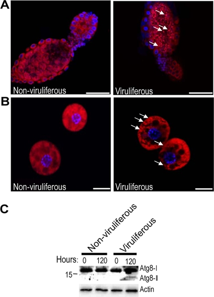
FIG. 6.
Detection of autophagy in adult whiteflies. Ovariole of ovary (A) and fat body (B) tissues stained with the lysosome-specific fluorescent dye Lysotracker Red (red) and the nuclear dye Hoechst 33342 (blue). Compared with the tissues from the nonviruliferous whitefly (left), there is increased punctate Lysotracker staining (white arrows) in the viruliferous whiteflies (right). Scale bar in panels A and B, 50 μm and 10 μm, respectively. Similar findings were observed in three independent experiments. (C) Western blotting of the Atg8 protein. Arabic numbers above each lane indicate the time points (in hours) after the nonviruliferous and viruliferous whiteflies were transferred to healthy cotton plants. A molecular size markers (in kilodaltons) is shown on the left of the panel. Atg8-I (∼16 kDa) is observed in all whiteflies, and Atg8-II (∼14 kDa) is induced only in the viruliferous whiteflies feeding on cotton plants for 120 h.

