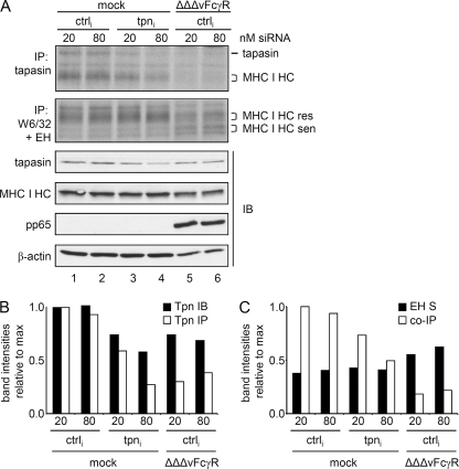
FIG. 5.
Blocked interaction between MHC I and tapasin in HCMV-infected cells. (A) MRC-5 cells were subjected to nucleofection with siRNA targeting tapasin transcripts or with a random sequence (ctrl) and then infected as indicated. Cells were metabolically labeled for 3 h prior to lysis at 48 h postnucleofection/postinfection. Lysates were analyzed by anti-tapasin (STC) or anti-MHC I (W6/32) immunoprecipitation (IP) or by immunoblotting (IB) with the indicated antibodies. (B) Diagram showing relative intensities of bands corresponding to tapasin (Tpn) IB (black bars) and IP (white bars). (C) Relative intensities of endo H (EH)-sensitive MHC I molecules (black bars) and tapasin-coprecipitated MHC I (white bars).

