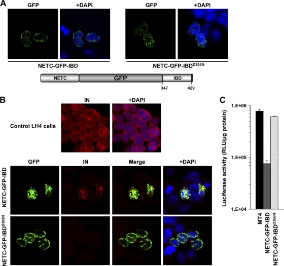
FIG. 7.
NETC-GFP-IBD inhibits HIV-1 infection. (A) NETC-GFP-IBD and NETC-GFP-IBDD366N display a nuclear envelope localization pattern. (B) NETC-GFP-IBD colocalizes with HIV-1 IN at the nuclear envelope, in contrast to NETC-GFP-IBDD366N, which does not. Cells were transiently transfected with the indicated constructs, and immunofluorescence analyses were performed as described in Materials and Methods. Untransfected LH4 cells are shown in the top panel for comparison. Note that IN is poorly visualized in LH4 cells in the presence of NETC-GFP-IBDD366N because it is not protected from degradation (28); the confocal gain is much higher in the control LH4 panel at the top than in the images of the bottom two rows, enabling the low level of IN to be seen in these LEDGF-deficient cells. (C) NETC-GFP-IBD and NETC-GFP-IBDD366N were stably expressed in MT4 with lentiviral vectors. The lines were challenged with HIV-1luc, and the luciferase activity was analyzed at 5 days.

