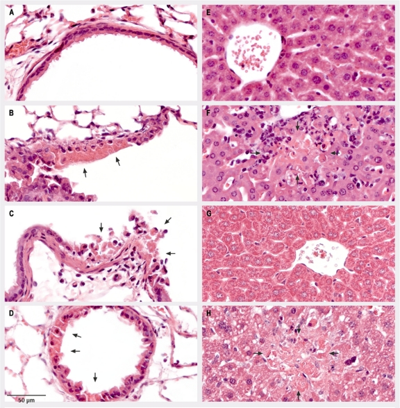
FIG. 4.
Histopathologic analysis of lung and liver tissues from C57BL/6 mice. C57BL/6 mice were mock infected (A and E) or infected intranasally with 200 PFU of the ECTV wt (B and F), ECTVΔN1L (C and G), or ECTV-N1Lrev (D and H). On day 6 p.i., thin sections of lung (A to D) and liver (E to H) tissues were prepared and subjected to staining with hematoxylin and eosin. Arrows indicate focal acute necrosis of epithelial cells of the bronchiolus (B, C, and D) and focal necrosis of hepatocytes (F and H).

