FIG. 4.
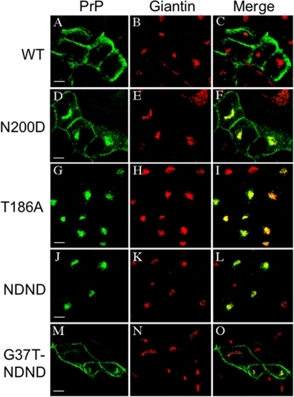
Accumulation of PrPC mutants in the Golgi compartment. PrP and giantin were labeled as for Fig. 2, and fluorescence emission was examined by confocal microscopy. Z-axis planes are shown. The mutants analyzed (D to O) are indicated on the left (for G37T-NDND, see the Fig. 3 legend). Bars, 10 μM.

