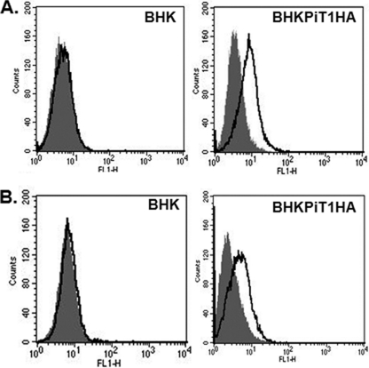
FIG. 1.
(A) PiT1 protein is expressed on the BHK cell surface. The expression of HA on the cell surface of BHK cells expressing PiT1HA was detected by flow cytometry after staining with Alexa Fluor 488-conjugated anti-HA antibody for 1 h (areas under bold line). Isotype controls are in shaded areas. (B) BHKPiT1HA can bind GALV envelope protein. V5-tagged GALV SU was used to bind BHK or BHKPiT1HA cells as described in Materials and Methods. FITC-conjugated anti-V5 antibody was used to detect GALV SU binding on the cell surface, as represented by the areas under the bold line; shaded areas represent isotype controls.

