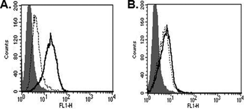
FIG. 7.
Binding of XMRV is heparin independent. (A) After CHOK1Xpr1 cells were treated with (areas under dotted line) or without (areas under bold line) heparinase II, the expression of HSPGs on the cell surface was detected by flow cytometry analysis after staining with anti-human heparan sulfate monoclonal antibody-FITC conjugate for 1 h. (B) Binding of XMRV envelope to CHOK1Xpr1 cells was determined with HA-tagged XMRV SU, similar to in Fig. 3. Areas under the dotted line show XMRV binding to CHOK1Xpr1 after treatment with heparinase II. Areas under the bold line represent XMRV binding to CHOK1Xpr1 without heparinase II treatment. Shaded areas represent isotype controls.

