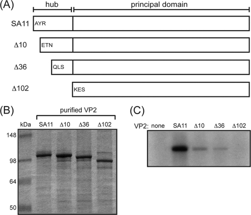
FIG. 5.
Truncation mutagenesis of the SA11 VP2 5-fold hub. (A) Cartoon schematics of wild-type and mutant SA11 VP2 proteins. The residues contributing to the 5-fold hub are delineated from those of the principal domain by a black line. The three amino acids that follow the starting methionine are listed for the wild type (SA11) and each mutant protein (Δ10, Δ36, and Δ102). (B) Purified VP2 proteins. VP2 proteins were electrophoresed in a 10% SDS-polyacrylamide gel and visualized by PageBlue staining. Molecular size markers are shown (in kilodaltons). (C) In vitro dsRNA synthesis by SA11 VP1. Reactions proceeded in the absence (none) or the presence of the different core shell proteins listed above the gel. Radiolabeled dsRNA products were resolved with 10% SDS-polyacrylamide gels and detected by autoradiography.

