FIG. 7.
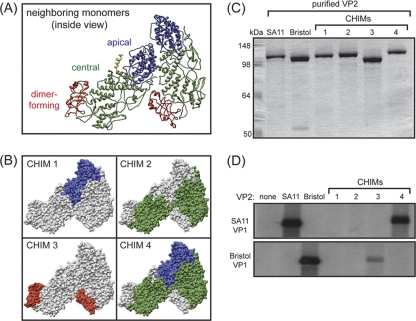
Subdomain chimeric VP2 proteins. (A) Ribbon representation of neighboring VP2 monomers in a dimeric unit (inside view). The apical, central, and dimer-forming subdomains are in blue, green, and red, respectively. The resolved portion of the VP2 amino terminus in the type B monomer (residues 81 to 100) is in yellow. (B) Surface representations of chimeric (CHIM) VP2 proteins. In all images, gray indicates Bristol VP2 residues, while color indicates SA11 VP2 residues. (C) Purified VP2 proteins. VP2 proteins were electrophoresed in a 10% SDS-polyacrylamide gel and visualized by PageBlue staining. Molecular size markers are shown (in kilodaltons). (D) In vitro dsRNA synthesis by SA11 or Bristol VP1. Reactions proceeded in the absence (none) or the presence of the different core shell proteins listed above the gel. Radiolabeled dsRNA products were resolved with 10% SDS-polyacrylamide gels and detected by autoradiography.

