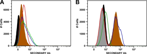
FIG. 1.
Staining of 220.221AEH cells and PBMC with DT9 and HLA-E-specific antibodies. (A) Staining of the 220.221AEH cell line, showing unstained cells (filled black area) and cells stained with MEM-E/06 (filled brown area), MEM-E/08 (red line), 3D12 (green line), and DT9 (blue line). (B) Representative staining of CD3+ lymphocytes (gating on appropriate forward and side scatter, live cells, and CD3 expression) from a healthy volunteer (HLA-A2, -B35, -B44, -Cw4, and -Cw5), showing unstained cells (filled black area) and cells stained with MEM-E/06 (filled brown area), MEM-E/08 (red line), 3D12 (green line), DT9 (blue line), and an IgG2b isotype control (filled gray area).

