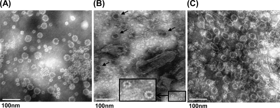
FIG. 5.
Electron microscope analysis of purified VLPs. Twenty microliters of VLP suspension was fixed in copper grids and negatively stained with 1% ammonium molybdate. Virus-like particles were visualized by using a FEI Tecnai G2 Spirit transmission electron microscope. (A) VLPs purified from insect cells by baculovirus. (B) Cell culture supernatant. BSRT7 cells were infected with rVSV-VP1, and cell culture supernatants were harvested at 48 h postinfection. The larger VLPs, 38 nm in diameter, are indicated by arrows. The smaller VLPs, 19 nm in diameter, are shown inside the box (with a magnified image provided). (C) VLPs purified from BSRT7 cells infected by rVSV-VP1.

