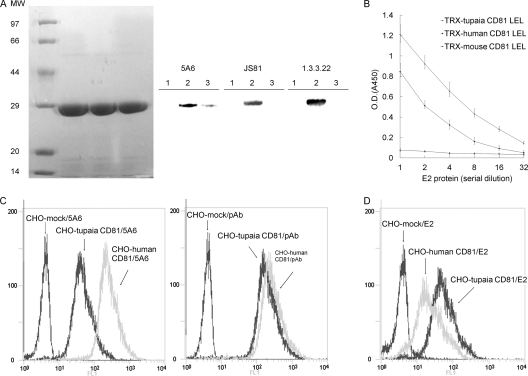
FIG. 2.
Tupaia CD81 binds HCV E2. (A) TRX-CD81 LEL fusion proteins were expressed and purified. Equal amounts of the fusion proteins were separated by SDS-PAGE and stained with Coomassie brilliant blue or immunoblotted with the anti-CD81 MAbs JS81, 5A6, and 1.3.3.22. Lane 1, TRX-mouse CD81 LEL; lane 2, TRX-human CD81 LEL; lane 3, TRX-tupaia CD81 LEL. MW, molecular weight (in thousands). (B) The interaction between HCV E2 and TRX-CD81 LEL fusion proteins was analyzed by EIA. Values are the means ± standard deviations of three independent experiments. O.D., optical density. (C) CHO cells were infected with lentiviruses containing human or tupaia CD81 sequences or mock lentivirus. Three days later, the cells were probed with the anti-CD81 MAb 5A6 (left) or polyclonal antibodies (pAb) (right) and analyzed for CD81 expression by flow cytometry. (D) HCV E2 binding to CD81-transduced CHO cells was detected with anti-E2 polyclonal antibodies by flow cytometry. In panels C and D, the values on the y axis indicate counts, and those on the x axis CD81 expression. The experiment was repeated at least three times, and one representative result is shown.

