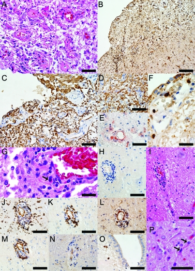
Figure 2. Neuropathologic findings in the medullary floor of the fourth ventricle and area postrema of the neuromyelitis optica (NMO) patient 1 illustrated in figure 1, A, C, and D.
(A) Thickening of the blood vessels in area postrema (hematoxylin & eosin [H&E], scale bar = 50 μm). (B) Profound activation of macrophages/microglia (KiM1P, scale bar = 100 μm). (C) Glial fibrillary acidic protein (GFAP) immunoreactive astrocytes extending foot processes that surrounded the endothelial cells are still present throughout these lesions (GFAP, scale bar = 50 μm). (D) GFAP immunoreactive astrocytes and preservation of neurons in the NMO area postrema (GFAP, scale bar = 50 μm). (E) Complement deposition in perivascular macrophages and astrocytes (C9neo, scale bar = 50 μm). (F) CD3+ T lymphocytes are present in the subependyma of the floor of the fourth ventricle (CD3, scale bar = 20 μm). (G) Eosinophils are components of the inflammatory infiltrates (H&E, scale bar = 20 μm). (H) Moderate perivascular inflammation in an area of aquaporin-4 (AQP4) loss (AQP4, scale bar = 50 μm). (I) Neurons are preserved around the blood vessel shown in H (H&E, scale bar = 50 μm). (J) Macrophages/microglia (KiM1P, scale bar = 50 μm), (K) CD3+ T lymphocytes (CD3, scale bar = 50 μm), (L) CD8+ T lymphocytes (CD8, scale bar = 50 μm), (M) B lymphocytes (CD20, scale bar = 50 μm), (N) Plasma cells (CD138, scale bar = 50 μm) are components of the perivascular inflammatory infiltrates. (O) Parenchymal plasma cells located in the subependyma of the floor of the fourth ventricle (CD138, scale bar = 50 μm). (P) Uninucleated, small, reactive astrocytes with thin, elongated processes (H&E, scale bar = 50 μm).

