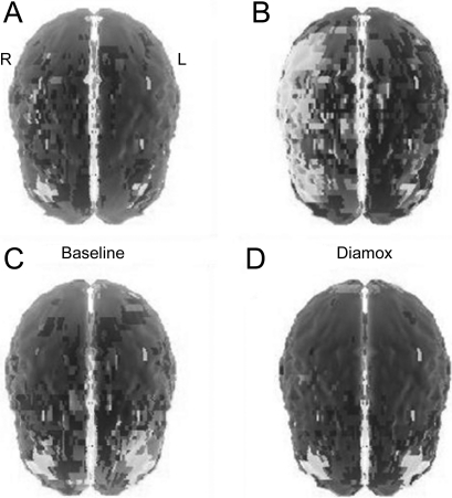
Figure 3. Assessment of cerebral perfusion.
SPECT scan (A, B) with acetazolamide (C, D) revealed decreased perfusion in the cerebral hemispheres, worse on the right, consistent with decreased vascular reserve in the middle cerebral artery territories (A, B) prestent, with a return of cerebral blood flow reserve (C, D) after stent placement.

