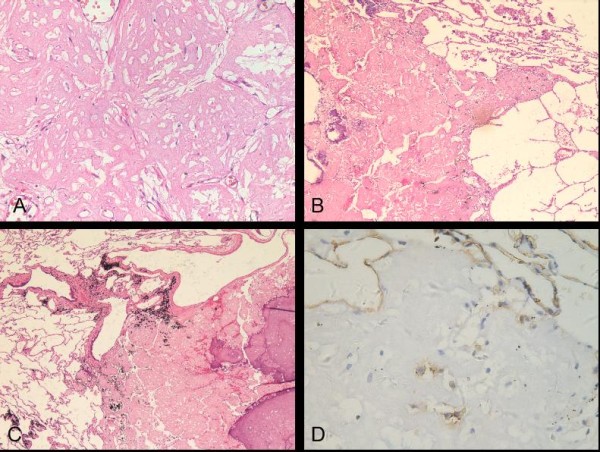
Figure 4.

Histologic findings of the multiple small nodules. (A) Eosinophilic substances were filled in pulmonary alveolar space; Cells in the nodule were small and moderate (H and E; original magnification ×200). (B) Border of the small nodule with partial calcification was not smooth and extended to neighbor airspaces (H and E; original magnification ×40). (C) The small nodule was located with the bronchiolar and vascular walls (H and E; original magnification ×40). (D) Individual small cells showed positive immunoreactivity for CD31 (original magnification ×400).
