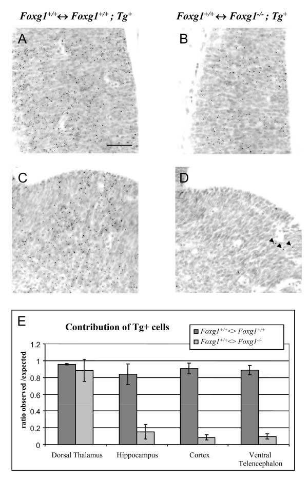
Figure 1.
Foxg1-/-mutant cells are underrepresented in the telencephalon of Foxg1+/+;Tg-↔Foxg1-/-;Tg+chimeras. (A-D) Coronal sections through the thalamus (A,B) and the ventral telencephalon (C,D) of E12.5 Foxg1+/+;Tg-↔Foxg1+/+;Tg+ control (A,C) and Foxg1+/+;Tg-↔Foxg1-/-;Tg+ experimental chimeras (B,D) showing Tg+ cells (labelled with dark dots) derived from the ES cells. (D) Very few Tg+ cells (arrowheads) are observed in the ventral telencephalon of experimental chimeras. Scale bar: 50 μm. (E) Ratios of observed/expected contributions of Tg+ cells in the dorsal thalamus, the hippocampus, the cortex and the ventral telencephalon of control (Foxg1+/+;Tg-↔Foxg1+/+;Tg+) and experimental (Foxg1+/+;Tg-↔Foxg1-/-;Tg+) chimeras. Tg+ cells are significantly underrepresented in the telencephalon of mutant chimeras (mean ± s.e.m, n = 3 embryos of each genotype; Student's t-test, P < 0.05).

