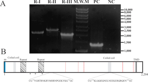
Figure 2.
cDNA amplification and PvRON2 schematic representation. A. PCR amplification from pvron2 gene RT-PCR product, with three sets of primers as described in the Materials and Methods section. Lane 1. pvron2 region I (~2,176 bp). Lane 2. pvron2 region II (~2,580 bp). Lane 3. pvron2 region III (~2,061 bp). Lane 4. molecular weight pattern. Lane 5. PvAMA-1 ectodomain amplification (positive control). Lane 6. Negative control. B. PvRON2 protein representation. The signal peptide is shown in blue, the transmembrane domain (TMD) in dark grey, coiled-coil motifs in light grey and red lines indicate conserved cysteines between Pf, Pv, Pk, Pc, Pb and Py. * represents polymorphic sites between Sal-1 (reference) and VCG-1 strains. The localization and sequence of inoculated peptides is marked.

