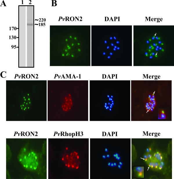
Figure 3.
PvRON2 expression and apical localization. A. Anti-PvRON2 rabbit polyclonal antibodies detected two bands at around ~220 and ~185 kDa in parasite lysate by Western blot. Lane 1: pre-immune serum. Lane 2: hyper-immune serum. B. P. vivax schizonts incubated with anti-PvRON2 polyclonal antibodies and revealed with FITC-labelled anti-rabbit IgG (green). Parasite nuclei were stained with DAPI (blue). C. Co-localization study: schizonts were simultaneously incubated with anti-PvRON2 and anti-PvAMA-1 (top) or anti-PvRhopH3 (bottom) and detected with FITC-labelled anti-rabbit and with rhodamine-labelled anti-mouse. Arrows indicate the typical dotted pattern displayed by apical organelles.

