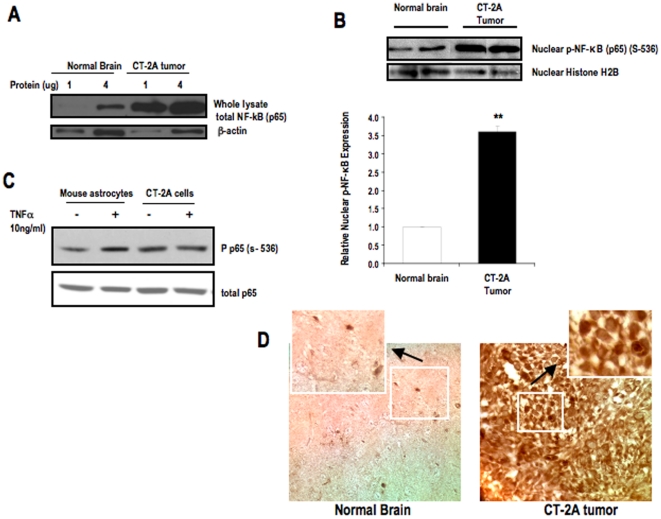
Figure 1. Expression and Phosphorylation of NF-κB (p65) in CT-2A astrocytoma.
Western blot analysis of (A) Total protein expression of NF-κB (p65) in the whole lysates of tumor tissue and normal brain parenchyma. 1 and 4 µg of protein was loaded for each sample of tumor and brain tissue. (B) Phosphorylated NF-κB (p65) in nuclear extracts of tumor and normal brain tissues. The histogram illustrates the average relative expression of p-NFκB (p65) (S-536) to histone in nuclear extracts of the indicated tissue. Values are expressed as normalized means ± S.E.M of 4–5 independent tissue samples/group for both A and B. The asterisks in indicate that the value is significantly higher in the CT-2A astrocytoma than in contra-lateral normal brain at ** P<0.01 (Student t-test). Two representative samples are shown for each tissue type. (C) Phosphorylation of NF-κB in CT-2A cells and control mouse astrocytes in absence and presence of TNFα (10 ng/ml). (D) NF-κB immunostaining in CT-2A tumor and contra-lateral normal brain tissues. 3 independent mouse brain tumors were analyzed.

