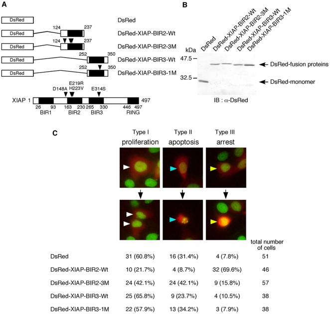
Figure 2. Overexpression of XIAP-BIR2, but not XIAP-BIR3, inhibits cell proliferation.
A) Diagrams showing the constructions of DsRed-XIAP-BIRs expression plasmids and the structure of XIAP with the positions of the point mutations. Mutated amino acid residues are indicated by arrowheads. B) Expression of DsRed and DsRed-XIAP-BIRs fusion proteins in 293T cells. 293T cells were transiently transfected with DsRed or each DsRed-XIAP-BIRs expression plasmid as indicated. At 18 hour after transfection, cells were harvested and lysates were subjected to immunoblot analysis with the anti-DsRed antibody. C) The effects of overexpression of DsRed-XIAP-BIRs on cell proliferation. HeLa-H2B-GFP cells were seeded at a density of 1×10⁵ cells per 35-mm glass bottom dish and incubated for 12 hours. Cells were transfected with DsRed or each DsRed-XIAP-BIRs expression plasmid as indicated and further incubated for 24 hours. Then, cells were observed with the time-lapse fluorescence microscope at 15-minute intervals for 24 hours (see Videos S1, S2, S3, S4, S5). The observation indicated that DsRed-positive cells were divided into three types of cells; type I, normally divided into two nuclei showing proliferation (white arrowheads); type II, condensed and fragmented nuclei showing apoptotic cells (blue arrowheads); type III, condensed but not fragmented nuclei showing proliferation-arrested cells (yellow arrowheads). The cell numbers of each type and the percentages to the total DsRed-positive cells are shown.

