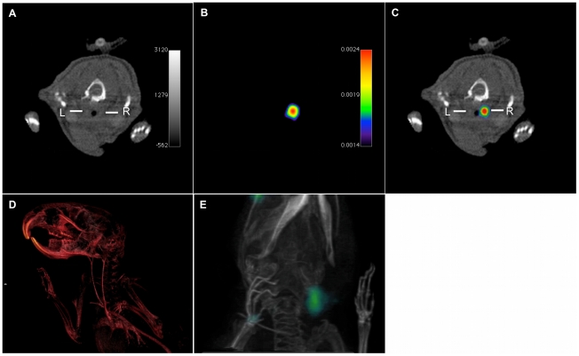
Figure 4. In vivo SPECT-CT imaging of carotid artery thrombosis.
In vivo SPECT-CT single (A, B) and fused (C) images after injury of the right carotid artery (right side) and incubation with 111In-labeled LIBS. CT angiogram of the neck region enhanced with iodinated contrast (Imeron 350) provides vessel contrast for anatomical detail of the carotid arteries (A, R = right carotid vessel, L = left carotid vessel); three-dimensional reconstruction of the CT-angiogram (D). SPECT imaging of the same neck region after incubation with 111In-LIBS depicts a co-localized peak of 111In-uptake (kBq/voxel). Three-dimensional overlay of CT data (D) with SPECT signal allows the correlation of the peak uptake to the area of the injured carotid artery (E) Movies of the three-dimensionally rendered images are provided as supporting information (Movie S1, Movie S2).

