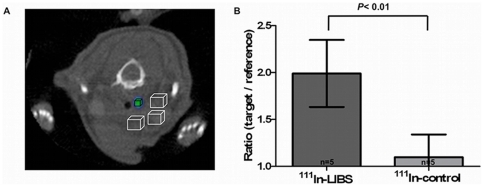
Figure 5. Evaluation of in vivo SPECT-CT results.
(A) SPECT images were evaluated with reference to the anatomical information provided by CT and based on the information gathered from histological sections about the location of the intravascular thrombosis. To compare the intravascular ligand uptake with the ligand uptake in the surgical bed, we defined 4 VOIs: A small VOI to best represent the target area of the thrombosis (mean 0.68 ±0.08 mm3; black box) surrounded by 3 reference VOIs (mean 3.63±0.3 mm3) with fixed location to assess ligand uptake in the surgical bed (cubes with an edge length of 1–2 mm; white boxes). From these VOIs we calculated a target to region ratio (i.e., mean uptake per volume in the black box divided by the mean uptake per volume of all white boxes). Comparing the ratio after injection of 111In-LIBS and 111In-control reveals a significant increase in ligand uptake after incubation with 111In-LIBS (B, P<0,01).

