Figure 4.
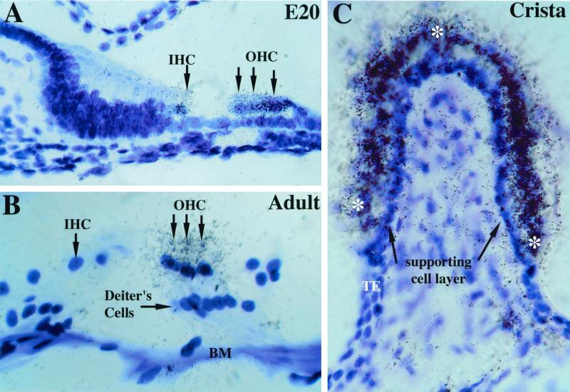
α10 nAChR subunit gene expression in cochlear and vestibular hair cells. (A) At embryonic day 20, both inner and outer hair cells (arrows) were decorated with silver grains after emulsion dipping of slides. No other structures in the cochlea were labeled (magnification ×630). (B) In adults (2–4 months old), α10 mRNA was detected only in outer hair cells and not in inner hair cells. BM, basilar membrane; IHC, inner hair cells; OHC, outer hair cells (magnification ×1,000). (C) Adult cristae express α10 transcripts as evidenced by the dense accumulation of silver grains over the entire surface (see asterisks) of the sensory end organ. Note the lack of positive reaction at both the transitional epithelium (TE) and the supporting cell layer (black arrows) underlying the hair cell layer (magnification ×630).

