
Fig. 1.
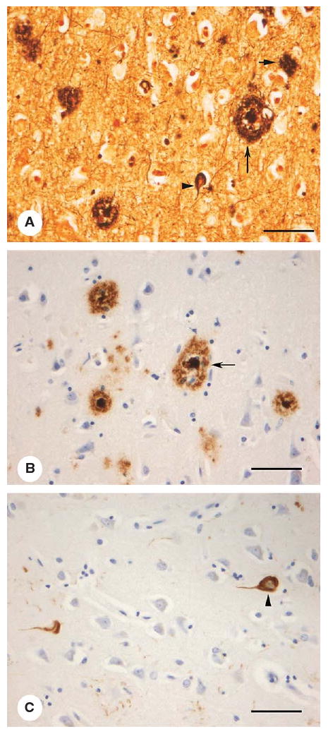
Histological images of AD-related lesions in neocortex: (A) modified Bielschowsky's silver stain, (B) Aβ immunohistochemistry demonstrating amyloid plaques and (C) hyperphosphorylated tau immunohistochemistry for neurofibrillary tangles. Senile plaques (SP, large arrow); diffuse plaque (DP, small arrow), and neurofibrillary tangles (NFT, arrowheads) (bar = 150 μm).