
Figure 6.
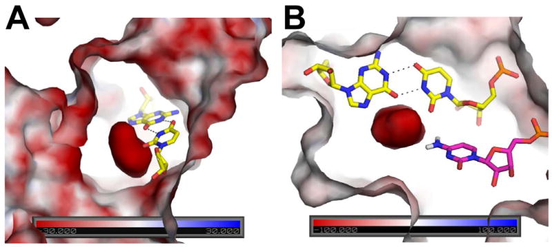
Surface electrostatic potential of precleaved HDV ribozyme. The potential is colored according to the scales provided in the base of each panel. Views shown here are near the reverse G•U wobble, which is depicted in yellow sticks with hydrogen bonding in black. (A) Orientation showing large negative potential in the center, which reaches values greater in magnitude than −100 kT/e. Analogous figure for the product state of the HDV ribozyme provided in Figure S5A. (B) Orientation showing interaction of C75 and reverse G•U wobble with negative potential in the precleaved state. C75 is in magenta with N4 hydrogens shown explicitly. Note that the C75 is oriented toward the highly negatively charged pocket generated near the reverse G•U wobble. The amine of C75 is a second shell ligand to the catalytic Mg2+ (not shown here) and is hydrogen bonded to the scissile phosphate. View is rotated counterclockwise by ∼90° from panel (A) and viewed from the top. Details of phosphates contributing to the potential of the precleaved state are provided in Figure S5B.