
Figure 1.
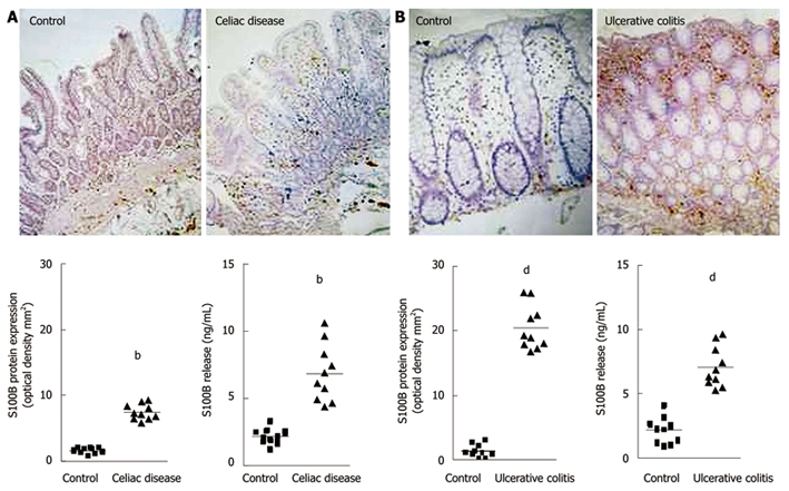
Changes in S100B protein expression during intestinal inflammation. A: Celiac disease[13]. Immunohistochemistry shows stronger S100B immunopositivity in the duodenal mucosa of patients affected by celiac disease, compared with healthy controls (original magnification, × 100). The graphs represent S100B protein expression (left) and release (right) in healthy controls and patients with celiac disease (bP < 0.01); B: Ulcerative colitis[14]. Immunohistochemistry shows stronger S100B immunopositivity in the rectal submucosa of patients with ulcerative colitis, compared with healthy controls (original magnification, × 100). The graphs represent S100B protein expression (left) and release (right) in healthy controls and patients with ulcerative colitis (dP < 0.01).