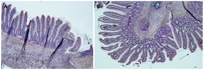
Figure 3.

Histological view. A: Chiu grade 0 with intact epithelium and a few villi along one side of the anastomosis (HE staining; magnification 5 ×), Region of the anastomosis (arrow) and staining artefacts (*); B: Chiu grade 2 with a pronounced subepithelial space in the villi next to the inverted anastomosis (arrow) and a suture hole (*) (HE staining; magnification 5 ×).
