
Figure 1.
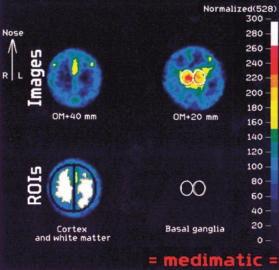
Upper row. Two imaging ‘slices’ 20 and 40 mm above the ear opening. The upper slice images the hemispheric cortex and the centrum semiovale. The lower slice images the basal ganglia/thalamus in the centre. Lower row: The regions of interest: lateral and mesial cortex, white matter, and basal ganglia/thalamus.