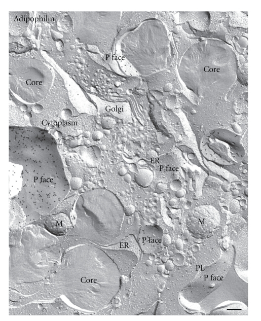
Figure 7.
Freeze-fracture overview of organelles in a lipid-laden macrophage after labeling for adipophilin. Gold particles marking the presence of adipophilin can be seen in abundance in the outer phospholipid monolayer (P face) of lipid droplets, ER membrane and plasma membrane (PL). The Golgi apparatus (Golgi) is devoid of label. M mitochondria. Bar: 0.2 μm.

