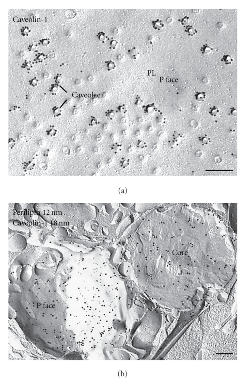
Figure 9.
Distribution of caveolin-1 and perilipin in adipocytes. (a) Caveolae appear as dimples in the P face of the plasma membrane of adipocytes. Gold particles label cavolin-1 in the P face at caveolae. Caveolin-1 labeling is found mainly at the rims of deep caveolae. Apart from positive labeling of caveolae in the plasma membrane, abundant label of caveolin-1 is found in the lipid droplet. (b) Example of immunogold labeling of perilipin (12 nm gold) and caveolin-1 (18 nm gold) in two lipid droplets of the same cell. One droplet shows colocalization of both proteins in the outer monolayer (P face) in almost equal amounts, whereas the other is cross-fractured and the core contains almost exclusively caveolin-1 label. Bars: 0.2 μm.

