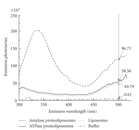
Figure 2.
Fluorescence resonance energy transfer to detect ATPase incorporation. Typical fluorescence emission from a single scan of liposomes (dotted) and proteoliposomes containing amylase (dashed) or ATPase (solid) and buffer (dashed dotted). All liposomes were prepared by filtration, contained 2 moL% dansyl, and were excited at 278 nm, the wavelength of excitation for tryptophan endogenous in the enzymes, and emission recorded over 300–520 nm; each preparation was scanned 3 times within 20 minutes. Mean emission intensities obtained from the three scans at 500 nm (grey vertical line), the wavelength of maximum emission of dansyl when excited by emitted light from nearby tryptophan, differed significantly within each replicate (a, b, c, d; n = 3).

