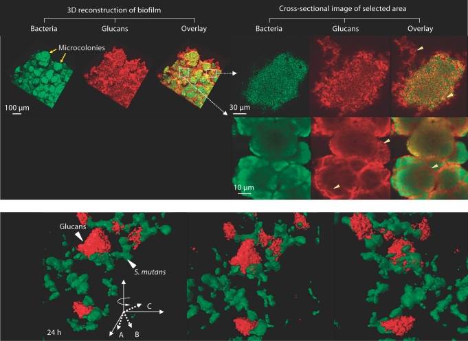
Fig. 1.
a Representative confocal images of bacterial cells (in green) and glucans (in red) within biofilms formed by S. mutans US 159 on tooth enamel surface in the presence of 1% (wt/vol) sucrose. Arrowheads indicate structural organization of glucans within microcolonies. b Close-up view of 3-dimensional structural relationship between glucans and S. mutans cells.

