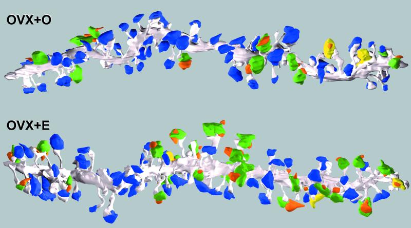
Figure 3.
Three-dimensional reconstructions of OVX + O and OVX + E dendritic segments. The dendrite is gray, SSBs are blue, different-cell MSBs are green, same-cell MSBs are yellow, and spines from other unfilled cells are orange. These are the last OVX + O and OVX + E dendrites in Tables 1 and 2. For the purpose of this figure, an MSB that made at least two synapses with the filled cell is shown as a same-cell MSB. Note the increase in dendritic spine and MSB density on the OVX + E dendrite, clustering of MSBs, and that the vast majority of MSBs are different-cell.

