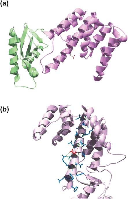
Fig. 1.
(a) Rv3124 structural model based on the EmbR molecular structure. The DNA-binding domain is in green, and the BTA domain in purple. Residue E159, located in the third α-helix of the BTA domain, is shown in red. (b) Close-up of the third α-helix of the BTA domain from a different angle. The side chains of residues that form the helix are in blue, except for E159, which is shown in red. Intra-chain H bonds are represented by green dashed lines. Replacement of Glu159 by Gly perturbs the helix.

