Fig. 3.
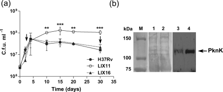
Effect of pknK deletion on growth of M. tuberculosis in culture. (a) Aerobic growth of H37Rv wild-type, LIX11 mutant and LIX16 complemented strain in 7H9-Tw-ADS medium was monitored for 30 days by measuring viable cell counts. The results presented are the mean±sd from three independent growth experiments. ** and *** represent P<0.01 and P<0.001, respectively, for the differences between the mutant and wild-type strain at a particular time point. Arrows indicate the time points, representing exponential phase (day 3) and late stationary phase (day 30), respectively, during growth at which samples were taken and analysed for PknK protein levels. (b) Western blot analysis of PknK protein in H37Rv cell lysates at days 3 and 30. The Ponceau S stained blot shows equal loading of total protein from day 3 and 30 samples (lanes 1 and 2, respectively). PknK production is induced in stationary phase (lane 4) compared to exponential phase (lane 3). M, molecular size markers.

